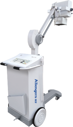
CONVENTIONAL X-RAY/ DIGITAL RADIOGRAPHY
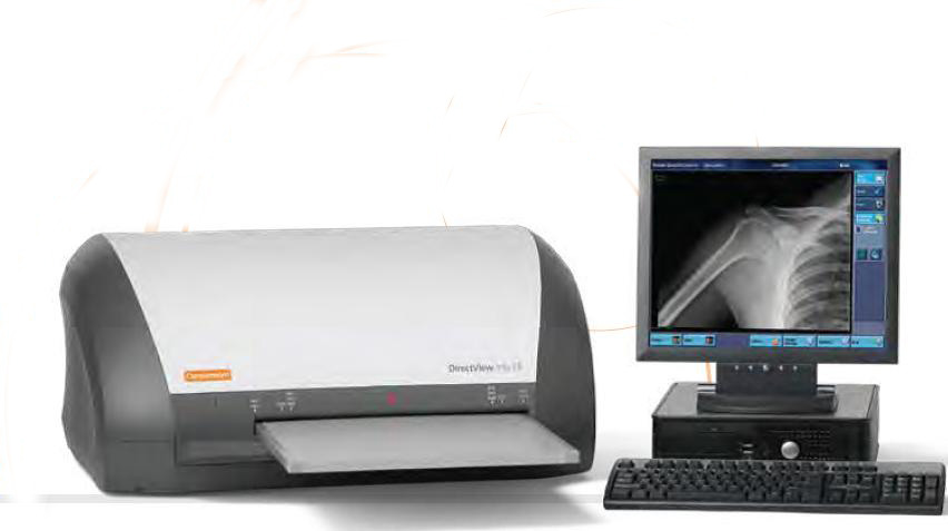
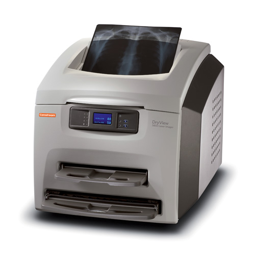
X-ray or radiography uses a very small dose of ionizing radiation to produce pictures of the body's internal structures. X-rays are the oldest and most frequently used form of medical imaging. They are used to help in diagnosis of fractured bones. look for injury or infection and to locate foreign objects in soft tissue. Some x-ray exams may use an iodine-based contrast material or barium to help improve the visibility of specific organs, blood vessels, tissues or bone.
Types of barium test:
- Barium swallow
- Barium meal
- Barium meal and follow through
- Barium enema
SONOGRAPHY
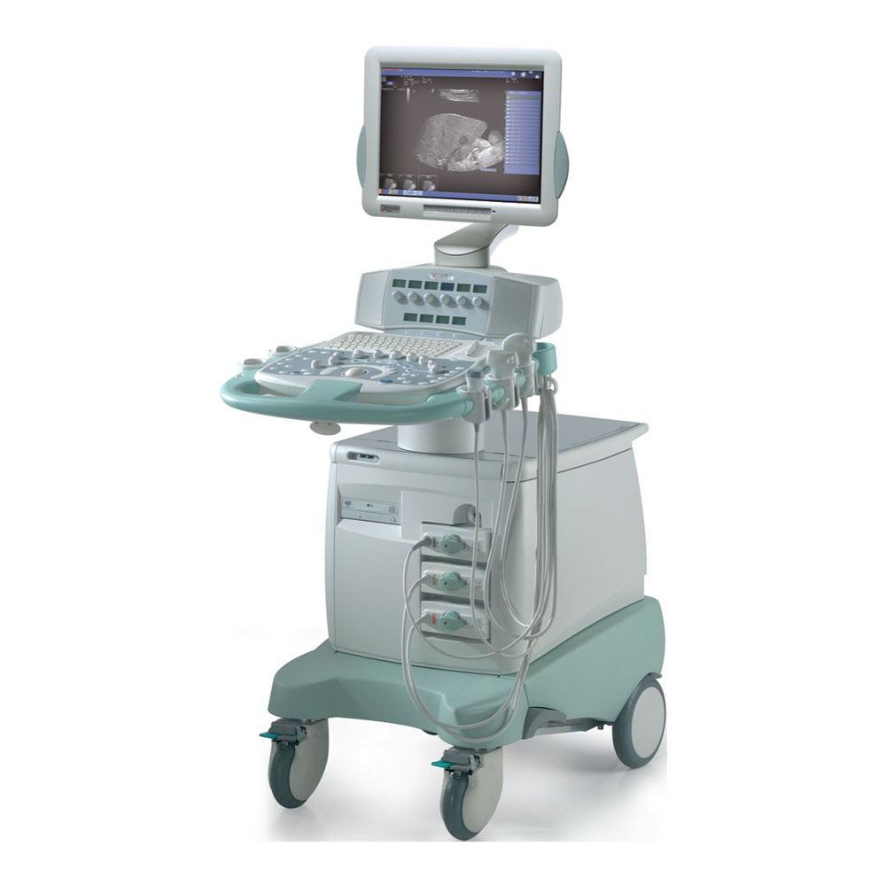
An Sonography scan or ultrasound scan is a medical test that uses high-frequency sound waves to capture live images from the inside of your body. An ultrasound allows to see problems with organs, vessels, and tissues-without needing to make an incision. Unlike other imaging techniques, ultrasound uses no radiation, so it is the preferred method for viewing a developing fetus during pregnancy.
It is safe and painless, and produces pictures of the inside of the body using sound waves. It involves the use of a small transducer (probe) and ultrasound gel placed directly on the skin. High-frequency sound waves are transmitted from the probe through the gel into the body. The transducer collects the sounds that bounce back and a computer then uses those sound waves to create an image.
Because ultrasound images are captured in real-time, they can show the structure and movement of the body's internal organs, as well as blood flowing through blood vessels.
CT SCAN
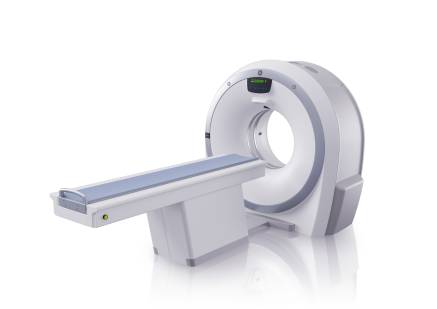
Computed tomography, more commonly known as a CT or CAT scan, is a diagnostic medical test that, like traditional x-rays, produces multiple images or pictures of the inside of the body.
The cross-sectional images generated during a CT scan can be reformatted in multiple planes, and can even generate three-dimensional images. These images can be viewed on a computer monitor, printed on film or transferred to a CD or DVD.
CT images of internal organs, bones, soft tissue and blood vessels typically provide greater detail than traditional x-rays, particularly of soft tissues and blood vessels.
Using specialized equipment and expertise to create and interpret CT scans of the body, radiologists can more easily diagnose problems such as cancer, cardiovascular disease, infectious disease, appendicitis, trauma and musculoskeletal disorders.
MRI SCAN
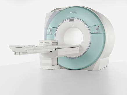
Magnetic resonance imaging (MRI) is a type of scan that uses strong magnetic fields and radio waves to produce detailed images of the inside of the body.
MRI is an imaging technique used primarily in medical settings to produce high quality images of the inside of the human body. MRI is based on the principles of nuclear magnetic resonance (NMR), a spectroscopic technique used by scientists to obtain microscopic chemical and physical information about molecules. The technique was called magnetic resonance imaging rather than nuclear magnetic resonance imaging (NMRI) because of the negative connotations associated with the word nuclear in the late 1970's. MRI started out as a tomographic imaging technique, that is it produced an image of the NMR signal in a thin slice through the human body. MRI has advanced beyond a tomographic imaging technique to a volume imaging technique.
MAMMOGRAPHY
Mammography is specialized medical imaging that uses a low-dose x-ray system to see inside the breasts. A mammography exam, called a mammogram, aids in the early detection and diagnosis of breast diseases in women.
An x-ray (radiograph) is a noninvasive medical test that helps physicians diagnose and treat medical conditions. Imaging with x-rays involves exposing a part of the body to a small dose of ionizing radiation to produce pictures of the inside of the body. X-rays are the oldest and most frequently used form of medical imaging
Three recent advances in mammography include:
- Digital Mammography
- Computer-aided Detection
- Breast Tomosynthesis
DEXA SCAN
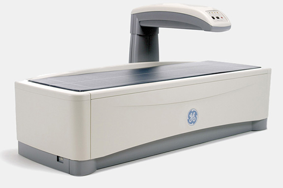
A dual energy X-ray absorptiometry (DEXA) scan, also called a bone density scan, is a common technique used to measure bone density. This completely painless procedure is easily performed and exposes the patient to minimal radiation.
During the scan, a large scanning arm will be passed over your body to measure bone density in the centre of the skeleton. As the scanning arm is moved slowly over your body, a narrow beam of low-dose X-rays will be passed through the part of your body being examined.
This will usually be your hip and lower spine - this is done to check for osteoporosis (weak and brittle bones). However, as bone density varies in different parts of the skeleton, more than one part of your body may be scanned. For some conditions, the forearm is scanned.